Thursday, 30 April 2020

Examination of Radial Nerve

2 questions that are relevant  to ask that are allowed in short case practical  examination are

(same holds true for all nerve related questions )

1. What happened ?

2. When it happened ?

if allowed ask

3. what are the difficulties for the patient

( Then introduce yourself , ask for permission to proceed and go to examination after proper examination )

Look- Scar

            Wasting of triceps , forearm radial borders


Feel - Temperature


           tenderness - Superficial and deep


         Tinel's sign - v. v.v. important

        check pliability of scar and scar tenderness, check tinel's on scar

          check bony union if there was a previous sign of fracture

          Lymph nodes

           Pulsation / Allen's test


Movement

           quickly assess upper extremity movements


Measurement
   

           quickly assess humerus shortening or  shortening of forearm


Neurovascular examination

Sensation

 Autonomous zone of all 3 nerves in UE

 Muscle power asessment

 Elbow Triceps

Forearm - Supinator , Pronator Teres , Brachioradialis

Wrist - ECRL , ECRB , ECU, PL , FCU , FCR


Hand

          Thumb

                EPL, EPB, Abductor Pollicis ,

           Finger

               EDC , FDS , FDP


Not to miss quickly  median nerve and ulnar nerve screening.

Reflexes 

Radial and Ulnar artery Pulsation important

Allen's test is important too .


This way all donor muscles are also tested quickly together while testing muscles supplied by radial

nerve so as to finish in time.


Donor tendons for radial nerve palsy are

          Pronator Teres

          PL , FCR , FCU

          FDS , 3rd and 4th finger


Deep Peroneal Nerve

First a brief about Common Peroneal Nerve

Two sensory branches

 1. Lateral Sural Cutaneous Nerve

      skin over lateral aspect of knee and proximal third calf

2. Branch that joins tibial anastomotic branch to form surnal nerve

     skin over posterolateral aspect of calf and over lateral malleolus , lateral aspect of foot , over 4th

     and 5th toes

Motor Supply

 1. Superficial Peroneal Nerve

    Motor ;     Peroneus Longus and Brevis

    Sensory;   Anterior and Lateral aspect of leg and dorsum of foot

2. Deep Peroneal Nerve

    Motor; Anterior Tibialis , Extensor Digitorum longus , EHL , Peroneus Tertius , Ist dorsal   


     interosseus

 (   Sensory; Ist web space via digital cutaneous nerve (lateral aspect of dorsum of great toe and

     medial  aspect of 2nd toe )


Treatment

   1.Splint

  2. ROM Exercises

 3. Repair - 60 - 70 % patients recover motor function .

      (nerve repair techniques  already discussed in previous section . )

4. Tendon Transfer

        Split Tibialis Posterior tendon
       

Sciatic Nerve Injury


Supply

Motor supply


Tibial Component

 Hamstrings , Gastocnemius , Posterior Tibialis , Long Flexor of Toes

Peroneal Component

 Anterior Tibialis , Long Extensor of Toes (deep peroneal nerve )

 Sensory supply

   Via Sural Nerve , Deep Fibular Nerve , Superficial Fibular Nerve , Medial Calcaneal Branches


Affects in

 Knee Flexion

 Ankle - Flexion / Extension

 Toe flexion / Extension

 Sensation - Loss

 Due to peroneal Divsion

         lateral aspect of leg , dorsum of foot

 Due to Tibial Division

          plantar aspect of foot


 Late deformities

 Equinus Deformity , Clawing of Toes , Atrophy of muscles supplied by this nerve.


Investigations

 EMG  for the Nerve

Investigation of the injury  - X-ray of the bone and joint to find out the cause.


Treatment

 Release comrpessing Leg cast


Endoneuroloysis for prolonged compression


Explore for fracture and dislocation of hip, shaft of femur fracture , knee dislocation early

            when no signs of recovery seen and if any chances of repair.


Penetrating Injuries on buttock -early exploration and repair


Methods of Closing gaps

  Knee flexion  , hip hyperextended - this method closes gap upto 15 cm

  resect part of femur.


Post operatively - Double spica cast

 6 weeks cast off , long leg brace with adjustable knee brace and gradually extend knee.


 ROM Exercises


Treatment of Brachial Plexus Injuries


How to proceed for the treatment ?


A. Open Brachial Plexus Injury

       Sharp Objects , Missiles injury


  Associated vascular and mediastinal injuries are treated first. Tag plexus nerves with suture.


  Document deficits

   EMG after 3 -6 weeks .


    Then Surgery.

      For  Low velocity injury

         wait and follow up in 3 and 6 weeks ,

          if no improvement then surgery .


B. Closed Injuries

       Traction injuries

        1.C5-6

        2. C 5-7

        3. C7 ,8,T1

  Observe , Physical Therapy  for 6 - 8 weeks.


    Myelography , axonal reflex evlauation

  Explore at 3- 6 weeks

                      if no functional recovery

                      if only distal recovery

                      post- ganglionic injury


Surgical Goals

     1. Restore elbow Flexion

     2. Shoulder Abduction

      3. Restore sensation to medial border of forearm and hand


Techniques


 Neurorraphy

 Primary Repair

 Neurolysis

Nerve Grafting

Neurotization


Direct intraoperative nerve stimulation of nerve axon potential obtained and Neurolysis  done.

If no nerve action potential obtained across damaged part , excision and grafting done.

If avulsion and no proximal stump found

          Intercostal nerve anastomosed with Musculocutaneous Nerve for Elbow Flexion .

          Takes 12-18 months  for nerve regeneration.


If patient presents late

Shoulder


            Tendon transfer

                         Trapezius to Deltoid transfer for Shoulder Abduction .


                L EPISCOPO Tendon Transfer

                              Latissimus Dorsi transfer for External Rotation of shoulder


                 Shoulder Arthrodesis if active scapulothoracic movement preserved.


   Elbow

                Flexion - Latissimus Dorsi , Pectoralis Major , Triceps , Sternocleidomastoid , Flexor   

                               Pronator mass tendon transfer .


Amputation -rarely for painful functionless limb.


Medial Cord Injury


Losses

A combined Median and Ulnar Nerve (except for FCR and Pronator Teres )

Extensive sensory loss

      medial aspect of arm and hand


Diagnosis

Clinical examination

 EMG including EMG of paraspinal muscles locates lesion in 80 % cases.


Posterior Cord Injury

Losses

 Motor Function

 Subscapularis Nerve function - Subscapularis and Teres Major 

Thoracodorsal Nerve - Latissimus Dorsi 

Axillary Nerve - Deltoid and Teres Minor 

Radial Nerve - Extensor of Elbow , Wrist fingers 


Sensory loss over Deltoid 


Functions affected 

 Shoulder IR / Abduction , Extension of elbow , Wrist and Fingers 


Improvement in triceps without deltoid suggests Axillary Nerve entrapment in Quadrangular Space.


Lower Plexus Injuries ( Klumpke )

Segmental sensory and motor deficits - C8 , T 1


       Motor loss - Intrinsic hand functions

                            Wrist and Finger flexors

       Sensory loss - over C8 , T1 dermatomes ( arm, forearm and hand )

Horner Syndrome


  Investigations
   

     Myelography

    EMG


Lateral Cord Injury

  Motor

 Musculocuataneous Nerve - Biceps weakness

 Lateral Root - Median Nerve - FCR , Pronator Teres weakness

Lateral Pectoral Nerve - Pectoralis Major

Glenohumeral Subluxation

Sensory Deficit

  anterolateral aspect of Forearm


Wednesday, 29 April 2020

Upper Brachial Plexus Injuries Investigation

After examination of brachial plexus  through our previous scheme of examining Brachial

Plexus , students have to know further detail about diagnosis and  management of different Brachial

Plexus Injuries. Its discussed below.


Upper Plexus Palsy (Erb's Palsy )

 C5-6     +/-  C7 Injury

Typical Position and  attitude of upper limb

         Elbow extended , shoulder Internally rotated , and adducted

         flacid rt side of trunk

Movement of Upper Limbs impairment
       
         Shoulder ROM - Abduction not possible

                                            due to paralysis of Deltoid and Supraspinatus

                                      External Rotation not  possible

                                            due to paralysis of Infraspinatus and Teres Minor

                                       Flexion  not possible

                                              due to paralysis of Biceps , Brachialis , Brachioradialis


            Forearm
                                         Supination not possible

                                           due to paralysis of supinator muscle


Neurological impairments


          Sensation absent over C5-6 myotome


          Paralysis of Long Thoracic Nerve and Dorsal Scapular Nerve leading to Winging of Scapula

       
Investigations

           Myelography

                   delay to 6 -12 weeks

                blood clot occlude opening to cord leading to pseudomeningocele

   CT with enhancements -overestimates root avulsion injury and dye extravasation if used early.

     MRI preferred

    Post Myelograph MRI and CT are mainstay of imaging in brachial plexus injuries.

Cutaneous Axonal Reflexes

          post ganglionic - no flare

          recovery possible after repair.


Upper Plexus after ruling out root avulsion exploration justified, repair sometimes possible.


Treatment part will be covered on next section.
   

Neurorraphy

Factors influencing regeneration after Neurorraphy


1.Age - higher chances of failing in elderly. Higher rate of success in children.


2.Gap between nerves - managed by nerve mobilization, transposition , joint flexion , grafting and

    bone shortening .

3. Delay in repair - upto 12 months - satisfactory results.

4. Level of injury - proximal muscles - good recovery.

                               proximal injury - incomplete motor and sensory recovery.

5. Conduction of nerve ends - for this expose fascicular bundles

6. Time of surgery -

                   primary repair - best done within 6-8 hours

                   delayed repair - best done in 7- 18 days.


Instruments required for neurorraphy

    Pneumatic tourniquet

     suction apparratus

      electrocautery

       Gelfoam and thrombin at nerve ends to control bleeding

       sutures ; 8-0 , 9-0 , 10-0 monofilament

        Epineural repairs ; 8-0 , 9-0

        Epiperineural repairs ; 9-0 , 10-0


Anesthesiae - regional , general or LA - Upper extremity

                        Spinal , general , LA - Lower extremity

Techniques


Exposure - Proximal and  distal ends

Endoneurolysis (Internal Neurolysis )

      if most fasciculi intact and separated and traced - nothing further done.


    If stimulation fails to illicit response , resect neuroma and neurorraphy done.


Partial Neurroraphy

     for large nerves e.g. Sciatic , Trunks, Cords of brachial blxus for partial severence

     suture - end to end ,

             if epineurium inadequate at injury site, epineural


Neurorraphy and Nerve grafting

       with gaps - median and ulnar nerve near wrist and elblow - nerve mobilization can be done.


         2-3 cm - Brachial plexus , Radial ,Sciatic ,Peroneal and Median nerves at midforearm level -

          they require nerve grafting.


Closing Gaps

           options

                        mobilization of nerve,  positioning of extremity, nerve transposition, bone resection ,

                        nerve grafting , neurotization

                        According to Zachory , median and ulnar nerves with gaps


                                                 upto 7-9 cm can be  fullfilled by   nerve mobilization


                                                   Anterior transposition helps closing gap as much as 13 cm .

                        According  to Spinner - not to stripe nerves more than 2-5 cm either side.

Tuesday, 28 April 2020

Management of Nerve Injuries

Following points to be included in one's scheme .


A. Detailed Clinical History

               1.      Particular detatils of patient

                            Hand dominance

                             Profession

            2.    C/C


           3.   History of Present illness

                  Regarding

                 Site of Injury

                  mechanism of injury -blast , crush , tractrion ,

                                                     open wound with contamination

                                                      weakness , progressing or improving

                                                     consequences of weakness on daily life

                                                    abilities related to the organ affected

             
                                                   constitutional symptoms

                       Any other injuries on body


       4. Treatment history -


                              Emergency management

                                         dressing  , debridement , tag sutures any ?


                            Definitive Management

                                         end -end anastomosis , end to soft tissue to prevent contraction

                                        coverage of soft tissues


                         PT               document use of dynamic or static splints

                                             Active or passive motion ?

                         Its outcome

                                              any contractures , infection  ,

                                              improvement in motor function

                                              improvement in sensation
                       
                                               improvement in functions


        5.  Past history

                 TT prohylaxis

                    drug allergy

                  DM and other medical illnesses


      6.   Expectation of Patient


B. Examination - Nerve examination mentioned in previous sections


C.Investigations

     1. Electrodiagnostic tests

          document injuries , location of inslt , severity of injury , recovery pattern , prognosis ,

        objective data for impairment documentation , pathology , selection of optimal muscles for

          tendon trasnsfers

  Nerve Conductions test (NCT ) and Electromyography (EMG )

         shows nerve conductivity , axon or myelin involvement  and muscles recruitment capability


Nerve Conduction test

        immediately after injury - interference of conduction but impossible to determine severity

        Proximal and distal to injury - Normal response

         Stimulation across injured segment varies

         5-10 days - Reduction in amplitude

                            - altered configuration of evoked potential

                          - Neuropraxia - conduction distal to lesion is normal .

       
Injury Pattern                     Porximal Stimulation              Immediately after injuries       after 10 days
                                          (conduction  distal to lesion)   (conduction across lesion )    (across lesion)

Neuropraxia                                  No response                       Normal response              Normal


Axonotemesis                            No response                            Normal                            No response
or neurotemesis

Proximal or central neurological lesions accessed using retrograde techniques (e.g. F-waves , SSEP )


EMGs
10-14 days after neural injury

 spontaneuous rest potentials + sharp waves appear in innervated myotomes with axonal injury


14 to 18 days

     fibrillation potentials

      denervation potential - last until innervated.


3 Weeks

            neural sprouting and increase in amplitude.

2 to 6 months

            large potentials remain until reinnervation completed.

Normal insertional activity present immediately after insertional activity.

Sharp waves


Amplitude - 100 - 400 microVoltage

 Duration 5 - 150ms

 Rate ; 2 - 40 Hz


wide body complexes

Fibrillation

Amplitude ; 5 - 1000 microvoltage
 Duration ;  0.5 - 2 ms

Rate;   2-3 Hz.


narrow body


onc can check the waves ;
https://www.researchgate.net/figure/Positive-sharp-waves-and-fibrillation-potentials-were-evident-in-all-four-limbs-upon_fig2_26881420

D.Treatment

Physiotherapy - ROM of the joints to prevent contracutre of joints

                           Splints

Nerve Repair -we will discuss later on different type of Nerve Repair. Upto now be able to examine

them properly from previous topics .

Tendon Transfer - be able to know different donor tendons  for different losses in different nerve

injuries more importantly in Upper Extremity. Radial Nerve examination and its losses is thoroughly

testested in examination.

Its equally important for median , ulnar nerves and brachial plexus injury.

Combined High Median and Ulnar Nerve

Anaesthesia over entire hand on palmar surfaace

Clawing of hand


Tr
Availability of Donor tendons for treatment

    Brachioradialis

     ECRL , ECRB, ECU , EIP


Omer

 1. Thumb arthrodesis

  2. Fingers - Zancoli capuslodesis - MCP joint of all fingers

 3. Release tendon sheath of flexor tendons

  4. ECRL to FDP (on radial border )

  5. Bachilradialis to FPL

  6. ECU to EPB on Ulnar side

  7. Amputate IF and fold radiallyinnervated dorsal flap into palm for sensation.

Combined Low Median and Ulnar Nerve

Complete anaesthesia over palm

loss of all intrinsics

loss of thumb functions

clawing - fixed if untreated


Tr

1. PT - to supple joints and skin

2. Finger intrinsics

                Brand transfer , ECRB (extended )


                Clawing  - Brown

                                   ECRL (extended ) to restore MCP as Brand .


  3.Thumb adduction

          EIP / FDS


          Omer

                    FDS LF- 4 tails / ECRL for adductor

   4. Opposition

          ECU extended by EPB or PL


    5. Fusion of Thumb for stability


      Options - ECRL for clawing , EIP for thumb adduction, ECU for thumb opposition to


          memorize for exam .


High Median Nerve Injury

Losses

  pronation of forearm

  flexion - IF, LF (Long Finger ) , thumb flexion

  opposition of thumb

  sensation over median nerve


Treatment


IF , LF -

     side to side anastomosis

          FDP 4th and 5 th finger

    or ECRL to FDP 2nd and 3rd finger


Thumb Flexion

       Brachioradialis

Opposition

       EIP

Sensation

       Neurovascular Island flap


Low Median Nerve Palsy

Deficits

Opposition of thumb

sensation over sensibility distribution

paralysis of 2 radial lumbrical muscles - little consequences with intact ulnar nerve


Functional requirement

Opposition of thumb

 Sensation on first web space - ? island flap thumb


Synergestic Muscles

Wrist Extensors

 FDS


High Ulnar Nerve Inujry

Same loss as low Ulnar Nerve palsy + FDP - RF and LF (additionally) .


Treatment

 Same as in low Ulnar Nerve Palsy but not to take FDS RF for treatment.

 FDP - 4th and 5th Finger

         side to side anastomosis with FDP 2nd and 3 rd finger

 Available  - wrist extensor , FDS , EIP.

Monday, 27 April 2020

Restoration of Intrinsic functions of fingers

Clawing

1.Stiles and Forrester - Brown

FDS - detach , split and transfer to dorsum of fingers to extensors

 No f/u


2. Bunnel

FDS - detach , split

   one slip to each of extensor aponeurosis by way of lumbricals

   effective when intrinsic only weak but not paralyzed ,


3. Fowler's modification

   FDS - splitted into 4 slips

    passed through volar side of deep transverse metacarpal ligaments , attached to radial side of extensor aponeurosis of each finger

effective if wrist is flexed.


4.Brand

ECRL / ECRB passed through volar side of forearm , to carpal tunnel and extended by 4 tailed grafts

Plantaris / Palmaris Longus ) and through lumbrical canal to extensor aponeurosis.


5. Riordan

FCR passed through dorsum of wrist, extended with 4 tailed graft , each tail passed volar to deep

transverse metacarpal ligament and attached to radial side of extensor aponeurosis .


6.Zancoli Arthrodesis

 capsulodesis , joint supple , no muscles for transfer .


7. Tenodesing

Riordan Technique

 -ECRB and ECU cut at M3/D3 portion on forearm , extended one slip of each with grafts and one

   slip left intact

-each slip altogether 4 extended into extensor aponeurosis passing volar to deep transverse

metacarpal ligament .


Restoration of Adduction of Thumb and Abduction of Index Finger


Adduction of Thumb

1. Boyes

Brachioradialis throgh 3rd interosseous space to dorsum  to thumb adductor tubercle along with

tendon graft of Palmaris Longus (PL ) or  Plantaris

Post-operatively - cast removal at 3 weeks and active exercises


2. Royle Thompson transfer (modified )

FDS ring finger transferred dorsoradially like Riordan , divided into two slips .


Distal one - appeoneurosis with EPL

Proximal slip - MCP joint capsule and Adductor Pollicis


Abduction of IF

1.EIP transfer

2. Abduction Pollicis Longus transfer

Restoration of Pinch and Opposition in Hand

A. Arthrodesis

  15 degrees IR , slightly flexed

   if no tendons for transfer , at 20 degrees flexion , IP joint for a IP joint flexion contracture .


B. Tendon transfers

 1 . Riorddan

   FDS RF through loop of FCU to thenar region to apponeurosis of EPL and Abductor Pollicis .


 Post - operatively - 3/52 dressing, then splint remove and active motion intermmitantly

     
 thumb splint in opposition for 6 weeks , teach opposition with RF.


2. Brand transfer

 FDS RF to thenar region  then split into two slips.

          1st  passed to ulnar side of thumb IP joint proximally

          2nd passed to Abductor Pollicis and EPL .


3. Bulkhalter

        EIP taken to subcutaneuos tissue and ulnar border of wrist to palm to thumb to MP capule


         thumb , Abductor Pollicis Brevis and EPL .


4. Grooves and Goldner

     FCU and Sublimus

      FCU - proximal portion alongwith sublimus passed and loop created by distal portion of FCU

       with ECU .


5. Camitz Technique

     PL to Abductor Pollicis Brevis

6. Muscles transfer , Littler and Cooley

 Abductor Digiti Minimi to resotre opposition

If no other muscles  , it also gives thenar atrophied muscles a bulk.


Pre-requisites for Tendon Transfer

Skin and joints supple

No bony malalignment

Restore sensation before tendon transfer

Polio - wait at least 18 months before surgery otherwise it may recur


         Meadin Nerve - wait for four months


Technical considerations -


Donor tendon should be pink and red

Poor nutrition - Pink and pale ; smaller than normal tendons.


Low Ulnar Nerve Palsy

Deficits

Pinch - aadductor Pollicis

            - Ist dorsal interossei

Grip - finger intrinsics


Treatment

Claw

A. Tendon Transfer


1.  RF/LF intrinsic paralysis

             EIP - 2 slips passed volar to deep transverse ligament , and passed to radial side of each

                         finger extensor finger aponeurosis

              Zancoli capsulodesis


2. Omer MCP thumb arthrodesed
 
             FDS RF 2 Slips made..
       
                                              1 slip passed across palm to fibres of adductor pollicis

                                              2nd slip further divided into 2

                                                     1  passed through lumbrical canal to radial side of extensor

                                                          aponeurosis RF

                                                     other to LF
           

           other technique

                                       Omer - Brachioradialis extended to adducor of thumb

                                       EIP radial half splitted and inserted to Ist dorsal interosseous muscle

                                     
3. Burkhalter Technique

          Brachioradialis /ECRL extended and taken to dorsum , volar to transverse metacarpal ligament

          then fixed to radial side of Proximal phalanx

4. Brown


          FDS RF deep to finger flexors

          EIP to palm through 3rd MC and across palm volar to deep transverse metacarpal ligament ,

           attach to radial side of each finger.

5.Bunnel / Brand transfer


B)  Rehabilitation


Sunday, 26 April 2020

Shoulder Examination

Look - Overlying skin

            Deltoid conotour

            muscles on supraspinatus and infraspinatus fossae ,

            inferior angle of scapula level same or not


Feel


  temperature

 Clavicle medial end , laterally , AC Joint , Acromion process , Spine of scapula ; medial border /


inferior angle and lateral bodrder of scapula , glenoid and humerus


Movement


 Flexion , abduction , extension , ER/IR both active and passive

measure the angle affected


Special Tests

Drop arm test on flexion and abduction


Cross arm adduction test

Adson's test

Hawkin's test


Neer's Impingement Test


Cuff Muscles

    Supraspinatus test

      Infraspinatus test

      Teres Minor test

      Subscapularis test

Apprenhension sign ,

Sulcus test

Tinel's sign on ulnar nerve and median Nerve


Radial , Ulnar ,Median Nerve  sensation test


Radial and ulnar artry pulsation


Test for ligamentous Laxity


D/D

C- spine pathology


Rotator Cuff Pathology

Compression Neuropathy #

Cubital Tunnel Syndrome

Carpal Tunnel Syndrome


Ulnar Nerve Examination

Not to miss following points during the examination

Attitute of the limb or hand

cascade of fingers


Look -
           any scars on arm , forarm

       guttering on the dorsum of hand , wasting on hypothenar region


Feel - temperature

           any soft tissue or bony tenderness

           tinel's sign ,scar tenderness , scar pliability

Movement

          active and passive

Measurement if any angulation or bony deformity


Neurovvascular examination

sensation of autonomous zones of hand and  direct the examination to the nerve affected

Power of muscles - Examine both the weaker muscles and donor group of muscles


  Elbow - Brachioradialis

             
Wrist      FCU


Hand       FDP - 4th and 5th finger , FDS Ring Finger , FDP 2nd and 3rd finger

               Lumbricals

              Opponens Digiti Minimi

              Abductor Digiti Minimi

             Adductor Pollicis

             FPB

Special tests to know the names .

    Card test - for Palmar interossei

     Igawa test- for dorsal interossei

     Froment's sign - flexor pollicis sucstitutes adductor pollicis

Reflexes

Pulsation / allen's test


How do you proceed ? 

detailed history , physical examination

NCS and EMG


What next ? 

Like any other nerve injuries ,see previous section on nerve injury management.


What are choices for tendon transfer  ? 

Low Ulnar Nerve Injury

          Brachioradialis

          EIP

         FDS from 2nd or 3rd finger


High Ulnar Nerve injury

         Brachioradialis

          EIP

          FDP from 2nd,3rd finger

          sensory flap


see the mnemonic

       BEF again like in median nerve


Examination of Radial Nerve and viva


Not to miss following points during the examination

Attitute of the limb


Look - Wrist Drop

             Elbow semiflexed

           any scars on arm , forarm

          any muscles wasting


Feel - temperature

           any soft tissue or bony tenderness

           tinel's sign ,scar tenderness , scar pliability

Movement

          active and passive

Measurement if any angulation or bony deformity


Neurovvascular examination

sensation of autonomous zones of hands and  direct the examination to the nerve affected

Power of muscles - Examine both the weaker muscles and donor group of muscles


          Arm - Triceps function


  Elbow - Brachioradialis

              Pronator Teres

             Supinator

Wrist      ECRL

             ECRB

           ECU


           PL / FCU / FCR


Reflexes

Pulsation

Special tests - ligament laxity


How do you confirm that its radial nerve injury ?

Humerus shaft fracture or dorsoradial fracture

 Deficits

           Wrist extension

            thumb extension and abduction

             finger MCP extension

More distal radial injuries
   
              low radial nerve palsy

              preserved wrist extension

             loss of thumb extension , abduction and finger MCP joint extension


Treatement -

Observe - closed humeral fracture


Surgery - in absence of recovery or no advancing tinel's sign at 3 months

                radial nerve  neurorraphy (outcome before 6 months )

                Tendon transfer - post pone for 6 months


Burkhalter indications

1.To act as a substitute during regrowth of nerve , avoids external splints

2. to act as a helper

3. to intervene when results of nerve repair poor or irrepairable

Low Radial Nerve Palsy

P3 forearm fractures


Different Tendon Transfer

Jone's -


Brand -


Boyes -


Tsugen -


Elbow Examination


Atttitude of the joint


Look - Around Joint and extremity for the skin , any muscles wasting on arm , forearm and hand


Feel - Soft tissue , any bony tenderness on medial condyle , tinel's sign on ulnar nerve , nerve palpation , Olecranon process , lateral condyle , pulsation of brachial artery

Feel axillary lymy nodes

Done all if history suggestive of Insatbility or old lateral condyle fracture otherwise a quick valgus / varus stress test done

Joint Stability test - Valgus stress test


Varus stress test -  with shoulder on internal rotaion

                               force varus done similar to valgus stress ,

                               avoid rotating limb when performing test


PLRI (Pivot shift test )

Lateral Ulnar collateral ligament insufficiency

manifestation - episodes of subluxation or dislocation

Position - UL -  Patient lies supine , shoulder flexed sothat limb lies above patient's head

                          examiner grasps  patient's forearm and ERof shoulder  done sothat forearm pronates

                          and elbow  extended.

Then Give Valgus and axial compression forces to elbow and a supination torque to forearm ,


It leads to Rotatory subluxation of Ulnohumeral joint with a coupled posterolateral dislocation of

radial head from humerus.

       
Movement

Flexion , extension passive and active

Measure Cubitus angle


Distal

All 3 nerves

Radial and Ulnar artery


Approach to management of nerve injuries

Detailed history and Physical Examination

Investigations

PT - for supple skin , with good  ROM of joints


Splinting


Neutotropic medications

Surgery

 not good after 9 months .


Thursday, 23 April 2020

Entrapment Syndromes

Median Nerve Entrapment (Pronator Syndrome )

Injury proximal to Elbow - Involvement of  Wrist , Fingers and Thumb 

Injury on proximal forearm -- Wrists spared 

Injury on Wrist - thenar group of muscles involved 


Benediction sigh - High median nerve injuries . 

D/D 
1. Bicipital Tendinitis 

     resisted elbow flexion illicits pain 

2. Resisted Pronation 

  with a finger 3 finger breadth below elbow crease illicits pain 


Radial Tunnel Syndrome 

PIN compression at Arcade of Forhse 

4 fingers below lateral epicondyle illicits tenderness. 

Resisted long finger  extension test  - finger and wrist at 30 degrees extension - give pressure to flex MCP passively. 

a severe PIN compression , ECU doesnot function and wrist goes into radial  deviation.  

Note 

Brachioradialis , ,ECRB , ECRL lie proximal to radial tunnel . 

ECU , EDC , EPL and EPB lie distal to Radial tunnel . 


Anterior Interosseous Nerve (AIN ) - 

Aching pain  simiilar to that of pronator syndrome . aching pain in proximal forearm. 

Weakness - FPL , FDP to IF and Pronator quadratus  - Ok sign +ve 
 

Cubital  Tunnel Syndrome 

Repititive movement,  RA , OA , Fracture , Dislocation , Cubitus Valgus , Instability , Anconeus Epitrochlearis crossing nerve in region of medial epidondyle 


Inspection - Clawing of fingers,  atrohpy of hypothenar muscles 

Palpation - Ulnar Nerve 
  
                   Tinel's sign - tip of long finger suggestive of irritation of nerve 

                                (radiating pain distally ) 

                    Instability - palpae elbow - flexion / extension 

                    Compression test 

                         holding more or  upto 1 min with elbow in maximum flexion leads to paresthesiae of                          
                           LF and RF 

                    
                     Finger abductioon / adduction (intrinsic weakness )

                      FDP - RF / LF weakness 

                      FDP - LF , RF and Wrist flexion in radial deviation 

                                   weakness suggestive of compression proximal to wrist. 

                      High lesion - ulnar clawing 


claw hand - weakend lumlbricals allow MCP hyperextension and it inhibits full extension of PIP     
                       
   joint . 


Bovier's test for claw hand - examiner passively prevents MCP hyperextension allowing PIP to fully extend. 


Investigation 

X-ray 

RBS 

TFT 

NCS , EMG 

USG / MRI 


Practical Approach to examination of Brachial Plexus

Brachial Plexus

Its a complex structure . Rembering all of its branches is difficult. Rembering further down to all

muscles supplied by each nerve is even more difficult.Finally the Brachial plexus site of injury can

be pin pointed just by examination .  Best way is to reharse with friends in the same order mentioned

as below. It makes examination fast , easy and simple.


Look

 Front
             Head at centre
 
             no ptosis , myosis , anhydrosis on forehead

             Shoulders - any scars  on shoulder ?
                           
            normal axillary folds

            Chest - wasting of Pectoralis Major

             No any fixed attitude of Upper extremities

 Side -     Normal overlying skin on shoulder, describe scars if present , UE
 
               Normal Deltoid contour , Biceps , triceps , forearm muscles ,

               no guttering on dorsum of hand , normal thenar and hypothenar muscles

              axilla clear


Back - Normal alignment of head , vertebrae

            both scpaula at same level ,

            normal posterior axillary fold ,
             
            describe scars which might be present on shoulder

            winging of scapula


Feel - Temperature , an mass on suprascapular region , soft tissue tenderness , deep bony tenderness ,

           scar - size , mobility , tenderness ,

Movement

             
        C- spine - flexion / extension


        Shoudler - Active / Passive

        Elbow - Supination / Pronation

                    flexion / extension

        Wrist - Flexion / extension

        Fingers - MCP joints - Active / Passive

                        IP joints - Active / Passive

  Lymph nodes

  Pulsation - radial and ulnar arteries

 Power

Back - shrugging for trapezius ,

           serratus anterior

            rhomboids

            subscapularis - Gerber's test

             Latissimus Dorsi


Front
          Pectoralis Major

          Supraspinatus

          Infraspinatus / Teres Minor (External rotator)


Sides - Deltoid , Biceps , Triceps , Supinator, Pronator  , Wrist extensor , Finger flexor


Your impression

Deficits - Pectoralis Major

                Deltoid

                 Rotator Cuff
   
                Latissimus Dorsi

                Elbow / wrist finger extensor

                Impaired C5-6 Sensation

Diagnosis is C 5-6 injury.

What level ?
 Posterior to clavicle .
Cause Pectoralis Major exits  below Clavicle.


Next Scenario

3 years old girl by birth

Deficits - Rhomboids ,
                 
                 rotator cuff muscles
             
               pectoralis major

                deltoid

                wrist and finger extensors

                 C 5-6 impaired sensation

Diagnosis is Erb's palsy


Erb's point
  C 5 nerve root

   C6 nerve root

    two divisions of uppper trunk

    dorsal scapular nerve

     nerve to subclavius


How to investigate ?

 X-ray Shoulder - AP / Lat . to see joint in position or not ?

CXR to see involvement of diaphragms

Electrophysiologic studies

                                                 NCS and EMGs
CT scan of Glenoid to see glenoid retroversion


Treatment

Early Presentation - Neurolysis for  lesion in continuity

Primary Repair

 Nerve Grafting

Neurotization with oberlin transfer

Late Presentaion

 tendon transfer

            Shah - Trapezius to Supraspinatus

            Release - Steindler

Osteotomy

            derotational osteotomy of humerus


NCS and EMG in Brachial plexus role - to be studied .


Practical Approach for the Examination of Recurrent Shoulder Dislocation

Look - from front side and back

            Alignment of Upper Extremity - Normal

            No fixed attitude

            Fullness anterior shoulder

           skin normal

           muscule wasting around shoudler - anteriorly , deltoid contour and supraspinatus and

            infraspinartus fossae

Feeling


          Temperature normal

          tenderness on anterior shoulder

           palpate axilla -axillary artry , lymph node palpable or not


Movement

          Abduction

          Adduction

         Felxion

         Extension

         ER

          IR

Special Tests

          Sitting position

                                Apprehension ,
                         
                                anterior drawer test

                                Sulcus test
          Lying  Position

                                 Jobe's relocation test

                                 Laxity test - Load and shift - anterior and posterior direction

                                         (supine , arm flexion and adduction )


                               Jerk Test

                                Circumduction test


Laixty test

Distal Neurovascular test


In summary - in sitting and lying positions


 tests for anterior instability

          Apprehension test and relocation test

          Load and Shift test

        anterior drawer test

  tests for posterior instabilty

       Posterior drawer test

       Load and shift

       Jerk test

      Circumduction test


In multidirectional instability , sulcus sign and laxity tests are positive.


Practical Examination of Torticollis

Examination from

Examination starts after consent and order from the examiner.

Expoure requires upto umbilicus .


Front

  bent on lt side ,
 
  chin deviated to  rt side

 decreased head shoulder distance on lt side

 oral cavity looks normal /abnormal

 no squinting

 facial asymmetry - normal ?

 lt side sternocleidomastoid - taut


chest - no muscle wasting

Side

   Ear almost touching shoulder  ,
 
   Deltoid contour normal

    elbow fully extended


Back

     b/l shoulder symmetrical , hairline

     scapula at same level

     occiput flat

     Thoracic and lumbar curvature maintained with no scoliosis


Gait and Squating - normal

 Feel - lt sternocleidomastoid taut , thick

           no tenderness on mastoid, sternum , clavice, AC joint , scapula , humerus and C-spine

          No palpable lymph nodes, no tenderness on supra / infraspinatus fossa

       
Movement

          Flexion / Extension , Rotation , Lateral Flexion both active and passive

          meaure the angle head is bent.

Vascular Examination

Neurologic examination of upper extremities  / lowe extremities

 dermatome / myotomes

 sensation

 Reflexes

Discussion on Klepel - Feil Syndrome , Sprengel's deformity , Causes of Torticollis and its treatmenT. It will be added in future posts.


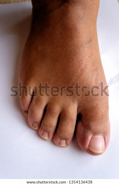
Hallux Rigidus

Rigidity of Ist MTP joint 

Gait - Altered 

Grerat toe straight , callus on medial side of distal phalanx 

MTP - Knobby , tender dorsal bunion 


outer side of sole - worn on footwears 

A HALLUX RIGIDUS ON FOOT


Treatement 

Not interfering activites - leave alone 

Intermittant pain 

       I/A injection , Local anaesthetics + corticosteroids 

       rock soled shoes 

      full length insole 


Surgery 
  
       chilectomy 

        Extension Osteotomy - PP 

        Arthrodesis 

                          10 degrees valgus and DF , 10 mm clearance 


       Arthroplasty 

       Interposition Arthroplasty  

     Capsular Arthroplasty 


         Metal implant better 

         silicone implant 


note - Taylor's Bunion oberlies 5th MT head. 


Deformities of Lesser Toes


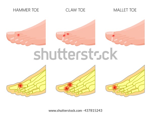
Claw Toes - MTP - Hyperextension

                    PIP -   Flexion

                    DIP -   Flexion


Hammer Toes - MTP - Extension

                          PIP - Acute Flexion

                          DIP - Straight

Mallet Toes -     MTP - Neutral 

                           PIP -  Neutral

                           DIP - Flexion


Illustration of the deformation of toes. Used: gradient, transparency, blend mode.

Rheumatiod Arhtritis -foot and ankle Clinical case


Attitude

Gait

Calf wasting

Clawing of  toes , swollen MTPs

Corns on dosum , plantar callosities

Skin - warm and tenderness

Non-fluctuant puffy swelling around ankle

movement restricted

Comment on adjacent joints


Lymph nodes - Popliteal and groin

Also comment on other joints on hands

DNVS

X-RAY  - Regional Osteoporosis

Blood Inv. - CBC ,ESR , RA - Factor , Anti- CCP antibodies

Biopsy

Treatement -

Special Shoes to accomodate toes

DMARDs

Synovitis - Corticosteroids , Operative Synovevtomy


Similar case may be present for Toe deformities

Note

Surgery for claw toes

Excision Arthroplasty for Hallux Valgus

TB Arthritis Ankle


TB Arthritis

Attitude

Gait

Calf wasting

Skin - wasrm and tenderness

Comment on adjacent joints

movement restricted

Lymph nodes - Popliteal and groin

DNVS

X-RAY  - Regional Osteoporosis , bone abscess , narrowing , irregularity of joint space

Blood Inv. - CBC ,ESR ,

Mantoux test

Joint fluid analysis

Biopsy

Treatement -

ATT

NWB in a caliper 

Wednesday, 22 April 2020

Dorsal SPINE TB MANAGEMENT BY MEHTA AND BHOJRAJ

According to  MEHTA AND BHOJRAJ 

Group A - Paradiscal and Central Involvement 

                    transpleural debridement with fusion (no instrumentation )


note - if Arthrodesis also required , then use transpleural approach instead of costotranseversectomy)


Group B - Group A + Deformity 

                    mx A + Instrumentation 

Group C  - to ill to undergo transpleural surgery 

                    transpedicular decompression and posterior instrumentation 


Group D - Posterior involvement only 

                 posterior decomression only 

Spinal TB Treatment and Approaches

If Deformity and disease activity both present then treat deformity as well as medically.


In general ,

Treatment

 Rest, Nutrition

 Chemotherapy

 Traction

  Surgery
Thoracic Spine -  Approach - A bit ambiguous, many options are there.

Anterior Transpleural ,Anterolateral  extrapleural and posterolateral


Posterolateral approach (Martin 1970 ) -Dura exposed by hemilaminectomy first and then operation

extended  laterally to remove posterior ends of 2-4 ribs, corresponding transevese process and

pedicles. He thinks anterolateral approach risky.


Approach to Atlanto-occipital and Atlanto-axial joint 

- numerous structures on the way anteriorly.

-supine - 5 to 10 degrees hyperextension , tracheostomy done .

Transoral Anterior Approach

- uvula, soft palate , bissected hypopharynx packed, 5 cm incision given and flaps raised. Apply stay

   sutures. Then anterior arch of atlas , body of axis and atlantoaxial joint exposed .

Anterior approach to spine 
Dorsal Spine - trasnpleural anterior approach (trasnthroacic / transsternal )

                         Extra-pleural Anterolateral (Costotransversesctomy )

Lumbar Spine - 
  
                          Retroperitoneal (Renal angle, Rectus Abdominis )

                         Posterior 

                          Transperitoneal (Linea alba , perotineum, bowel )

Posterior approach - Errector Spine -


                                                  Superior - Lateral Dorsi

                                                  Deep

                                                             Superior -sacrospinalis
                                                             Deep  - multifidus and rotators


parts of human ribs = head , neck , body , angle , costal groove , tubercle for articulation with vertebra


History taking of a traumatic patient in ward

Name - Muhammad Ashad                         Age / Sex - 9 yrs / m  wt. - 20 kgs

Gr II stduent , Lahore,  Pakistan               History Teller - Father

Site of taking history  - Orthopaedic Dept., XYZ Hospital , Lahore , Pakistan

Date of examination - 1 July, 2017

C/C - Pus discharge left leg with limping gait for 18 months

HOPI - M. Ashad was involved in RTAin Jan 1 , 2016. He was a pilon rider  and  hit by a bus from

the side and fell down. He had a shear injury on the leg and foot. Bike rider did not have any major

injury and walked immediately. Ashad had unbearable pain on foot and leg with a large wound on

dorsum of the foot, shin and lateral side of the leg.Leg was deformed with bone exposed on the shin.

He was soon taken to Emergency Dept., ABC Hospital in Gujrawala,Lahore within 45 minutes in a

taxi with leg wrapped using a handkerchif. He was in shock when they reached the hospital.He had

no LOC, ENT bleeding and vomiting . Left leg was bandaged to control bleeding after dressing .

Bloodwas arranged.III pint of colloid was given.  II pint of B + ve group whole blood  was given

there after recieving blood. Back slab was applied upto mid-thigh. After stabilization over 6 hours he

was referred to XYZ hospital.Patient reached Emr Dept. XYZ Hospital in next 6 hours. He was

further resuscitated with IV fluids.There he underwent x-ray evaluation for PRIMARY TRAUMA

SERIES which were normal.FAST-scan was normal. Hb was 9.0 gm %. X-ray of the left leg

inculding knee and ankle was done which showed shaft of tibia / fibula fracture. X-ray of the foot

was normal. He was diagnosed as Gu IIIb Shaft of Tibia/ Fibula fracture with Degloving injury of

left  leg on calf , shin  and foot with Shock.He was given Inj. TT 1 ampoule im stat.Then  he was

admitted in ICU for 2 days. Wound irrigation and lavage followed by dresssing was done in ICU. He

was given Inj. Gentamycin 100 mg iv .od , Inj. Cefazolin 150 mg iv tds. , inj . ketolak 5mg iv tds.

His lab reports (CBC, LFT,RFT, Urine R/e , RBS , Viral markers for HIV I and II , Hepatitis B and C

) were normal.Then patient  underwent serial  wound irrigation and lavage followed by dressing for

next 2 days in ICU with IV midazolam 5mg.

In Jan 3 , 2016 he underwent Skin Grafting and external fixator application with skin donation from

opposite thigh. He was transferred to ward in post-operative period.The outcome was uneventful

during course of treatment. Skin graft was uptaken on 5th day.He had no any post- operative

complication.He was discharged on 7th post- op day. External fixator was removed after 6 weeks

visit in Feb 21,2016 in theatre and discharged same day with back slab. Slowy pin tracts healed.

In April 1,2016 (12 weeks after injury ) he was admitted and  underwent IMILfixation for Shaft of

tibia fracture next day electively.It was also uneventfull. IV medications (Inj. Cefazolin and

Gentamycin , Ketolak ) were given.On 7th day he was discharged.

3 months after surgery (IMIL ) pus discharge was distally on medial side on the leg in follow up

which is persisting.He had no fever. ESR was raised to 30. He was advised to do dressing daily.

Now he has limping gait which is progressive. His left leg is shorter which is worsening .He walks

around 1 hour and then feels tired. He can walk up and down the ladder with the support on the side

bar. He has no difficulty using toilets.He has no fever.He sleeps well .His appetite is normal and

gaining weight.

Past History - No known drug allergy ; no known illnesses in the past.

Family Hx. - 5 memebers in family. 2 brother and 1 sister. They have a 1 storoyed building. His

father is a bus driver.


Personal Hx. - Student of Gr II . He is non-vegeterian.

Birth and Dev. Hx - He has normal growth and developmental milestones.

Vaccination hx. He has completed all the vaccines as per EPI schedule.


Expectation -Limb length Equlalization and go to school .


Based on History - Diagnosis is

 Chronic Osteomyelitis Lt Tibia with Limb Length inequlity as a sequelae of post traumatic lt shaft

of tibia / fibular fracture with IMIL in situ


Flail Limb

Different Options

Free Functional Transfer

lateral dorsi or Nerve to  Pec Major to lateral extensor mass

Extraplexal transfer
 
      Cross C 7 to Suprascapular nerve

      Phrenic Nerve to  c 5,6


       Intercostal to C 7


Management priorities

  Elbow flexion
 
  shoulder abduction

Discussion Supracondylar Fracture of Distal Humerus

Age 6- 10 years

common in boys.

Fall on outstretched hand (FOOSH) - common mechanism of injury.

Break in periosteum anteriorly , hinge force posteriorly.


Extension fracture in 95 % of cases
Flexion type fracture in 5 % of cases .

Extension type fracture classification of Gartland and Wilkin's

1. Undisplaced fracture

2.Displaced fractures , posterior cortex intact

3.Displaced fracture , posterior cortex breaks


Flexion type
direct injury on elbow on flexed position.


Patient presentation

1. history of trauma

2.pain , swelling , deformity , bruises on arm

3.Deformity - S-shaped , puckering ,

4.compratment syndrome

( It may be due to vessel spasm , contusion , tear , thrombosis
If capillary refill present , hand arm Dunlop traction given. )

5.Nerve injuries
 proximal fragment spike hits the nerve.
type 3a posteromedial displacement - radial nerve injured

 type 3b with posterolateral displacement -  ulnar nerve affected.

flexion type injury results in Ulnar nerve injury


X-ray description

AP film
  s/c fracture , fracture line oblique or transverse through olecranon fossa

   displaced or undisplaced

Lateral  - fracture displacedment direction , fat pad signs


Tr.

1.Type I - Slab / LAC

2. Type II - Slab / LAC
 
     Medial column communition - pinning

3. Pinning /Open

Reduction - realign coronal tilt

                    push anteriorly to correct extension .

X-ray assessment

Lateral Landmarks
posterior margins of coronid fossa
anterior margins of olecranon fossa

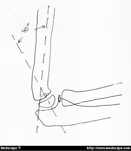
  Shaft condylar angle
  angulation of 40 degrees between the long axis of humerus and long axis of lateral condyle

Supracondylar and Lateral Condyle Fractures of the Humerus in ...


Humeral ulnar angle - best resembles the carrying angle of elbow .


AP Film

Bauman's angle - 64 to 81 degrees
Radiography of the elbow supracondylar fracture. Baumann's angle ...


https://www.google.com/search?q=baumans+angle+in+supracondylar+fracture+royalty+free+images&tbm=isch&ved=2ahUKEwib7rWjlPzoAhXZFHIKHVqEA8MQ2-cCegQIABAA&oq=baumans+angle+in+supracondylar+fracture+royalty+free+images&gs_lcp=CgNpbWcQA1C55xhY3-8YYIj3GGgAcAB4AIABuQSIAfUPkgEHMi02LjUtMZgBAKABAaoBC2d3cy13aXotaW1n&sclient=img&ei=KkmgXpuxIdmpyAPaiI6YDA


Open reduction indications
failure of closed methods

NV compromise

open fractures

Pining Types
Most stable - Cross Pinning

Lateral - Divergent

               Convergent - less stable

              parallel - least stable , toggles

Wilkin's Recommendation. - 2 divergent pins from lateral side ; of them one pin high in shaft leads to most stable fixaiton . He is a proponent of lateral pin entry.